Portable CMOS x-ray system enables Harvard's lizard evolution research
Researchers at the Losos Lab at Harvard University’s Department of Organismic and Evolutionary Biology and Museum of Comparative Zoology—which focuses on the behavioral and evolutionary ecology of lizards—are studying the competitive effects of a species invasion with the goal of testing the criteria for ecological character displacement. Led by Jonathan Losos, PhD, the lab’s major questions concern how Anolis lizards, commonly known as anoles, interact with their environment and how anoles have diversified. The team is studying the evolutionary response of Anolis carolinensis, the only native anole to Florida, to the invasion of A. sagrei from Cuba. Since their introduction, they have spread across Florida, presenting a novel competitor to the native A. carolinensis.
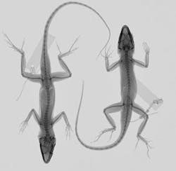
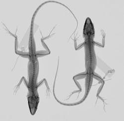
Researchers needed specific criteria—such as skeletal changes—to conclude that differences between species actually constitute character displacement due to competition. Seeking a portable x-ray imaging system to screen Anolis lizards in the field, with the idea of studying whether A. carolinensis was evolving its morphology in response to competition with A. sagrei, Harvard graduate student Yoel Stuart began working with Rad-icon Imaging (Sunnyvale, CA, USA) and its partner Kodex (Nutley, NJ, USA) in 2009 to assemble a portable CMOS x-ray imaging system, with the goal of catching anoles, imaging them on site, and then returning them to their natural habitat. Stuart did not want to alter the natural experiments by taking individual lizards back to Harvard’s more traditional x-ray machine.
System requirements included high resolution, cost efficiency, and an active area large enough to image the subject, but not so large to add additional cost. Harvard’s imaging capabilities had been limited only to preserved specimens that could be brought back to the university. The system at Harvard included a 130-kVp micro-focus x-ray source, a Varian (Palo Alto, CA, USA) PaxScan 4030R amorphous silicon detector, and a veterinary table with a movable tubestand. This imaging setup required animals to be euthanized or preserved.
Preliminary imaging was performed using an Oxford Instruments (Abingdon, UK) Apogee x-ray source at 40 kVp, with 0.1-mm AL filter, 1-mA source current, 1-s exposure time, 1-m distance to source, and 20-frame average rate. Two specimens were placed within the detector active area directly on the camera cover plate. The results allowed the team to move forward with specifying the system.
In May 2009, Rad-icon began to prepare a Remote RadEye200 camera system for Harvard. The total imaging system included a Remote RadEye200 with a 14-bit Shad-o-Box camera module with GigE adapter. The Ethernet interface connects to a Dell laptop with a 2-Mpixel display for fast image download.
Kodex integrated the x-ray camera with a power supply, aluminum stand, and 50-kVp portable x-ray source from Source-Ray (Bohemia, NY, USA) with a digital interface in GUI. Some components were mounted with Velcro to be easily dismounted and carried in a backpack for easy traveling in the field. The x-ray source mounted directly on top of the stand, yielding a 50-cm source-to-detector distance. The x-ray source and camera are not triggered together; images are controlled via Rad-icon’s ShadoCam software.
“Our new imaging system works great,” says Stuart. “Now we can collect crisp, clear images of bones in our lizards, which enable us to study evolutionary change much more precisely. This is absolutely necessary for us since A. carolinensis and A. sagrei have only been evolving together for a short amount of time, and we expect the amount of evolutionary change to be small.”
-- By Alana Achterkirchen, Rad-icon Imaging, a division of DALSA Corp.
-- Posted by Carrie Meadows, Vision Systems Design


