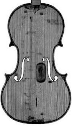
CT imaging helps reproduce Stradivarius
Using computed tomography (CT) imaging and CNC machining, a team of experts has created a reproduction of a 1704 Stradivarius violin.
Dr. Steven Sirr, a radiologist at FirstLight Medical Systems (Mora, MN, USA), worked with professional violin makers John Waddle and Steve Rossow of St. Paul, MN, on the project with the aim of producing an accurate reproduction of the original that could be played by young musicians.
To do so, the original violin was scanned with a 64-detector CT scanner, after which more than 1000 CT images were converted into stereolithographic files that could be read by a CNC machine.
The CNC machine, custom-made for the project by Rossow, then carved the back and front plates and scroll of the violin from various woods. Finally, Waddle and Rossow finished, assembled, and varnished the replica by hand.
For owners of authentic Stradivarius or other prized violins, CT imaging not only provides a definitive form of identification, it helps establish a pedigree that may increase the value of their investment.
"CT is useful in measuring wood density, size and shapes, thickness graduation, and volume measurements," Sirr says. "It also provides detailed analysis of damage and repair."
Three-dimensional images of the valuable violin and details on how the replica was made were presented at the annual meeting of the Radiological Society of North America (RSNA).
-- By Dave Wilson, Senior Editor, Vision Systems Design
