Cell-CT scanner may help to diagnose cancer faster
An imaging system under investigation at the Biodesign Institute atArizona State University (Tempe, AZ, USA) may help researchers pinpoint subtle aberrations in the nuclear structure of cells, leading to an improvement in the time it takes to diagnose cancer.
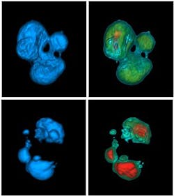
A team led by Professor Deirdre Meldrum examined normal, benign, and malignant cells using aCell-CT instrument from VisionGate (Phoenix, AZ, USA) that is capable of imaging cells in vivid 3-D with true isotropic resolution. The technology permits the examination of subtle cellular details inaccessible by more conventional forms of microscopy that are inherently 2-D.
Cell-CTimages cells in 3-D using a technique called optical projection tomography. It operates much like a normal CT scanner, though it uses visible photons of light rather than x-rays. Cells prepared for observation are not placed on slides but are instead suspended in gel and injected through a microcapillary tube.
As the capillary spins, the cell is scanned from multiple perspectives yielding a set of pseudo-projection images. These images are combined together using a method known as filtered back-projection, producing a final 3-D cell volume. Movies of cells seen in rotation reveal shape asymmetries, a particularly useful tool for disease diagnosis.
Currently, the definitive clinical diagnosis of malignancy relies on careful examination of the nuclear structure of cells that have been prepared by histological staining and subjected to brightfield microscopy. To do so,pathologists qualitatively examine cell features including nuclear size, shape, nucleus-to-cytoplasm ratio, and the texture of cell chromatin. However, these observations do not involve quantitative measurements that would promote a more accurate analysis.
The researchers at the Biodesign Institute say that the subtle nuclear differences observed using the Cell-CT system -- particularly with regard to the malignant cells -- would likely have been missed had the samples been examined with conventional imagery.
The results they have obtained using the system so far indicate that the system may dramatically improve 3-D nuclear morphometry, leading to a sensitive and specific nuclear grade classification for breast cancer diagnosis.
-- By Dave Wilson, Senior Editor,Vision Systems Design
