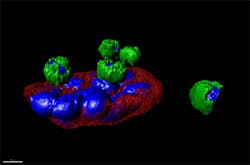
Imaging helps team to detect cancerous cells
Scientists from The Scripps Research Institute (La Jolla, CA, USA) have demonstrated the effectiveness of a blood test for detecting and analyzing circulating tumor cells (CTCs) -- breakaway cells from patients’ solid tumors -- from cancer patients.
The findings show that the highly sensitive blood analysis provides information that may soon be comparable to that from some types of surgical biopsies.
The new test, called HD-CTC, labels cells in a patient’s blood sample in a way that distinguishes possible CTCs from ordinary red and white blood cells. It then uses a digital microscope and an image-processing algorithm to isolate the suspect cells with sizes and shapes unlike those of healthy cells.
Just as in a surgical biopsy, a pathologist can examine the images of the suspected CTCs to eliminate false positives and note their morphologies.
“It’s a next-generation technology,” said Scripps Research Associate Professor Peter Kuhn, senior investigator of the new studies and primary inventor of the high-definition blood test. “It significantly boosts our ability to monitor, predict, and understand cancer progression, including metastasis, which is the major cause of death for cancer patients.”
The researchers’ studies were published in the journal Physical Biology and summarized here by Professor Kuhn.
-- by Dave Wilson, Senior Editor, Vision Systems Design
