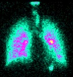
3-D imaging helps lung disease sufferers
Researchers at Southampton University (Southampton, UK) are using a 3-D imaging technique that could improve the lives of those suffering from chronic lung disease.
The research, led by Joy Conway, Professor of Inhalation Sciences at the University of Southampton, aims to provide a better understanding of diseases such as chronic bronchitis, cystic fibrosis and asthma.
The diagnostic imaging technique combines the data from a gamma camera and CAT scanner, after which a 360º image of the lung is created. The technique allows the researchers to examine how a particular drug is inhaled, dispersed and exhaled from the lungs.
The image can then be used to inform experts on how best to optimize the administration and delivery of inhaled drugs such as antibiotics and anti-virals for diseases like asthma, but also future gene therapies for diseases such as cystic fibrosis.
It is hoped the images will also assist physiotherapists in the future, helping to improve the effectiveness of chest physiotherapy required by many patients as part of a daily routine to help clear secretions from the lungs.
-- by Dave Wilson, Senior Editor, Vision Systems Design
