Image sensor detects pH
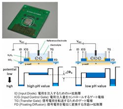
A newimaging sensor developed by Kazuaki Sawada from the Toyohashi University of Technology (Toyohashi Tech; Toyohashi, Aichi, Japan) not only enables researchers to capture images of cells in solution but also the pH as well.
The CMOS device consists of an array of CCDs that have been combined with a filterless fluorescence detector that can be used to detect changes in the concentration of hydrogen ions. In addition to monitoring the pH distribution, the device also yields optical images of the test sample.
The sensitivity of the pH imaging sensor enables the pH of the solution to be determined with an accuracy of +/- 0.0001 pH. "High sensitivity is possible because we accumulate charge over well-defined periods of time," says Sawada.
The current pH image sensors consist of 128 x128 pixels, each with a sensing area of 10 x 25 micrometers. But Sawada and his group are now developing pH image sensors with one million pixels, with each pixel being 10 x 10 micrometers.
Sawada said that in the future, he hopes to develop pH imaging devices for visualizing the movement and distribution of ions including calcium and sodium.
Sawada's group has recently reported on the use of the sensor for real-time imaging of acetylcholine (ACh) enzyme reactions. “We imaged changes in the distribution of ACh when nerve cells are stimulated with KCL,” says Sawada. Insights in the variation of the concentration of ACh may lead to new methods for the treatment of Alzheimer’s disease.
Sawada and his group are now looking for industrial partners to help put the pH image sensor to use in commercial instrumentation.
More information is available here.
-- by Dave Wilson, Senior Editor,Vision Systems Design
