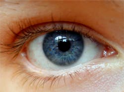
Images of the eye linked to heart disease
A pioneering study aims to determine whether a scan of blood vessels in the eye can identify signs of heart disease.
More than 1,000 patients with suspected heart disease will have high definition images taken of their retinas to check for indications -- such as changes to blood vessel widths or unusually branched blood vessels -- that may be linked to heart disease.
The study could find a way to identify whether a patient is at risk of heart disease, without the need to carry out invasive procedures such as biopsies or angiograms, where catheters are used to identify vessel and organ damage.
The project is led by the University of Edinburgh’s Clinical Research Imaging Centre (CRIC; Edinburgh, UK) and is a collaborative initiative with the University of Dundee, NHS Lothian’s Princess Alexandra Eye Pavillion, NHS Tayside’s Ninewells Hospital and Moorfields Eye Hospital in London.
The researchers will use specialist equipment from Optos (Dunfermline, UK) to see whether CT scans are a more efficient, cost effective and less invasive alternative to current procedures to detect heart disease.
“We know that problems in the eye are linked to conditions such as diabetes and that abnormalities in the eyes’ blood vessels can also indicate vascular problems in the brain. If we can identify early problems in the blood vessels in the eyes we might potentially pinpoint signs of heart disease. This could help identify people who would benefit from early lifestyle changes and preventative therapies,” says Dr. Tom MacGillivray, a research fellow at the University of Edinburgh and manager of the Image Analysis laboratory in CRIC.
-- by Dave Wilson, Senior Editor, Vision Systems Design
