Diagnostic imaging tool detects peripheral arterial disease
A new optical imaging tool created by Columbia University (New York, NY, USA) researchers may soon make it easier to diagnose and monitor peripheral arterial disease (PAD), one of the most serious complications of diabetes.
PAD, which is marked by a narrowing of the arteries caused by plaque accumulation, frequently results in insufficient blood flow to the body’s extremities and increases a person's risk of a heart attack or a stroke.
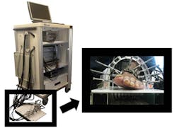
The new noninvasive imaging technique -- known as dynamic diffuse optical tomography (DDOT) imaging -- uses near-infrared light to map the concentration of hemoglobin in the body’s tissue.
Recently, the researchers revealed how they have developed a DDOT imaging system to reconstruct two-dimensional cross-sectional images showing hemoglobin concentration within the foot.
To map the presence of hemoglobin, the foot is illuminated with near-infrared light from laser diodes coupled into multimode fiber bundles. The light is then absorbed or scattered as it propagates through the foot, depending on the concentration of hemoglobin. It is then collected by detector fiber bundles positioned around the foot, after which it is analyzed to produce an image.
"Since hemoglobin is the main protein in blood, we can image blood concentrations within the foot without using a contrast agent," explains Professor Andreas Hielscher, the director of the Biophotonics and Optical Radiology Laboratory at Columbia University. Contrast agents pose the risk of renal failure in some cases, so the ability to monitor PAD without using a contrast agent is a great advantage.
The Columbia researchers hope to bring their diagnostic tool to market and into clinics within the next three years.
A recent paper on the imaging system entitled "Dynamic diffuse optical tomography imaging of peripheral arterial disease," which was published in Biomedical Optics Express, can be found here.
Related articles on diabetes from Vision Systems Design that you might also find of interest.
1. Smart phone application manages diabetes
An interdisciplinary research team at Worcester Polytechnic Institute (WPI; Worcester, MA, USA) has received a $1.2 million award from the National Science Foundation to develop a smart phone application that will help people with advanced diabetes and foot ulcers better manage their disease.
2. Hyperspectral imaging helps diagnose ulcers
Researchers at the UCLA Henry Samueli School of Engineering and Applied Science (Los Angeles, CA, USA) have developed algorithms that can analyze hyperspectral images of the foot to identify ulcers associated with diabetes weeks before they are visible to the eye.
-- Dave Wilson, Vision Systems Design
