Imaging system gets under the skin
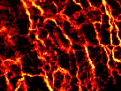
Researchers from Medical University Vienna (MUW; Vienna, Austria) and the Ludwig-Maximilians University (Munich, Germany) have used optical coherence tomography (OCT) to noninvasively image the network of blood vessels beneath the outer layer of skin, potentially revealing telltale signs of disease.
The researchers tested their system on a range of different skin conditions, including a healthy human palm, allergy-induced eczema on the forearm, dermatitis on the forehead, and two cases of basal cell carcinoma, the most common type of skin cancer on the face. Compared to healthy skin, the network of vessels supplying blood to the tested lesions showed significantly altered patterns.
"The condition of the vascular network carries important information on tissue health and its nutrition. Currently, the value of this information is not utilized to its full extent," says Rainer Leitgeb, a researcher at MUW and the study's principal investigator.
Ophthalmologists have used OCT since the 1990s to image different parts of the eye and the technology has recently attracted increased interest from dermatologists. OCT has many advantages over other imaging techniques. It is non-invasive and provides high-resolution images at high speed.
OCT is typically used to show tissue structure, but it can also reveal the pattern of blood vessels, which carry important clues about disease, by capitalizing on the unique optical properties of flowing blood cells.
The team's images of basal cell carcinoma showed a dense network of unorganized blood vessels, with large vessels abnormally close to the skin surface. The larger vessels branch into secondary vessels that supply blood to energy-hungry tumor regions. The images, together with information about blood flow rates and tissue structure, could yield important insights into the metabolic demands of tumors during different growth stages.
The research was published in the Optical Society's (OSA) open-access journal Biomedical Optics Express. The researchers' paper -- In-situ Structural and Microangiographic Assessment of Human Skin Lesions with High-speed OCT -- is available here.
Recent articles on optical coherence tomography (OCT) from Vision Systems Design.
1. Imaging system sees bacteria behind the eardrum
A new medical imaging device invented by a research team led by University of Illinois (Champaign, IL, USA) electrical and computer engineering professor Stephen Boppart allows doctors to see biofilms behind the eardrum to better diagnose and treat chronic ear infections.
2. Spectroscopy aids in characterizing skin cancers
A joint US-Dutch team under the direction of Anita Mahadevan-Jansen from Vanderbilt University (Nashville, TN, USA) has developed a noninvasive probe capable of both morphological and biochemical characterization of skin cancers.
3. OCT provides nondestructive testing at high resolution
Engineers at Santec (Aichi, Japan), have used National Instruments LabVIEW FPGA module to prototype the design of a portable OCT system.
-- Dave Wilson, Senior Editor, Vision Systems Design
