Telerobotic system treats bladder cancer
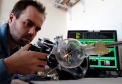
Engineers and doctors atVanderbilt University (Nashville, TN, USA) and Columbia University (New York, NY, USA) have developed a prototype telerobotic system that can provide surgeons with a better view of bladder tumors so they can diagnose them more accurately before removing them.
The machine itself is the size and shape of a large Thermos bottle but its end is only 5.5 millimeters in diameter and consists of a segmented robotic arm. The arm can curve through 180 degrees, allowing it to point in every direction including directly back at its entry point. At the tip of the arm is a white light source, an optical fiber laser for cauterization, a fiberscope for observation and tiny forceps for gripping tissue.
The researchers report that they can control the position of the snake-like arm with sub-millimeter precision: a level adequate for operating in clinical conditions. They have also demonstrated that the device can remove tissue for biopsies by gripping target tissue with the forceps and then cutting it off with the laser.
The fiberscope produces a 10,000-pixel image that is directed to a digital video camera system. Because it is steerable, the instrument is able to provide closeup views of the bladder walls at favorable viewing angles. However, the testing revealed the camera system's effectiveness was limited by poor distance resolution. According to the researchers, this can be corrected by re-designing the fiberscope or by replacing it with a miniature camera tip.
In the future, the researchers intend to incorporate additional imaging methods for improving the ability to identify tumor boundaries. These include a fluorescence endoscope, optical coherence tomography that uses infrared radiation to obtain micrometer-resolution images of tissue and ultrasound to augment the surgeon's natural vision.
More details on the system can be found here.
Related items from Vision Systems Design that you might also find of interest.
1.Artery walls imaged in vivo
Optical imaging technology from Wasatch Photonics (Logan, UT, USA) creates images of coronary artery walls in vivo to show where lesions and plaques have formed.
2.Kinect system helps doctors review records
Doctors may soon be using a system in the operating room that recognizes hand gestures as commands to tell a computer to browse and display medical images of the patient during a surgery.
3.MRI system images muscles in 3-D
Researchers at the Technische Universiteit Eindhoven (TU/e; Eindhoven, The Netherlands) and the Academic Medical Center (AMC) in Amsterdam have developed a technique that allows muscle structures to be imaged in 3-D.
4.Hand held instrument inspects the retina
Medical technology firm Lumetrics (Rochester, NY, USA) has won a $973,000 grant from the National Institutes of Health (Bethseda, MD, USA) to fund the development of a digital hand-held diagnostic ophthalmic instrument for inspecting the human retina.
-- Dave Wilson, Senior Editor,Vision Systems Design
