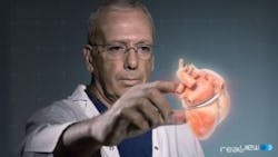
Clinical study shows 3D holographic images of the heart during procedures
Royal Philips and RealView Imaging Ltd. have completed a clinical study in which they demonstrated the ability to use 3D holographic visualization and interaction technology to guide minimally-invasive structural heart disease procedures.
The studies were conducted on eight patients in collaboration with the Schneider Children's Medical Center, where RealView Imaging’s visualization technology was used to display the 3D holographic images acquired by Philips’ interventional X-ray and cardiac ultrasound systems, according to a press release.
RealView’s technology enabled physicians to view a patient’s heart on both a 2D screen and as a detailed holographic image floating in free space during a procedure. Physicians can manipulate the 3D projection by touching the holographic volumes in front of them. This technology, in theory, could potentially enhance the context and guidance of structural heart repairs.
"The holographic projections enabled me to intuitively understand and interrogate the 3D spatial anatomy of the patient's heart, as well as to navigate and appreciate the device-tissue interaction during the procedure," said Dr. Einat Birk, pediatric cardiologist and Director of the Institute of Pediatric Cardiology at Schneider Children's Medical Center, in the press release.
The results of these clinical trials were presented at the 25th annual Transcatheter Cardiovascular Therapeutics (TCT) scientific symposium, sponsored by the Cardiovascular Research Foundation on October 29. The goal of the study, according to the press release, is to deliver innovative solutions that enhance clinical capabilities and improve patient experience and outcome. RealView and Philips will continue to explore the capabilities of 3D imaging and medical holography, both in this format and in other clinical areas.
This isn’t the only thing we’ve seen as of late, when it comes to the use of advanced imaging technology and visualization in the medical setting. Take a look here at five recent examples of innovative medical imaging techniques.
If you’ve seen something else we should know about, please let us know in the comments section below!
View the press release.
Also check out:
Carnegie Mellon to develop robots for bridge inspection, surgery
Augmented vision system aids the sight impaired
How robots could remove blood clots with medical imaging and steerable needles
Share your vision-related news by contacting James Carroll, Senior Web Editor, Vision Systems Design
To receive news like this in your inbox, click here.
Join our LinkedIn group | Like us on Facebook | Follow us on Twitter | Check us out on Google +
About the Author

James Carroll
Former VSD Editor James Carroll joined the team 2013. Carroll covered machine vision and imaging from numerous angles, including application stories, industry news, market updates, and new products. In addition to writing and editing articles, Carroll managed the Innovators Awards program and webcasts.
