High-speed imaging technique reveals mechanics of insect flight
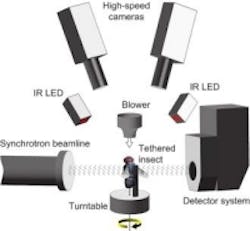
Researchers from various European academic institutions have developed an imaging technique that uses a particle accelerator, high-speed cameras, infrared LED lighting, and a detector system to apply time-resolved X-ray microtomography, which provides 3D visualizations of a blowfly’s internal movements.
In an open-access study published on PLOS Biology, it is explained that Dipteran flies are amongst the smallest and most agile of flying animals, and that their wings are driven indirectly by large power muscles, which cause cyclical deformations of the thorax that are amplified through the intricate wing hinge. In order to visualize the movements and deformations, the researchers developed a technique that uses a particle accelerator to record high-speed X-ray images of the flying blowflies, which were used to reconstruct 3D tomograms of their flight motor at ten different stages of the wingbeat.
Two synchronized Photron SA3 cameras with 180 mm Sigma macro lenses were used to film the blowflies, recording at 4,000 Hz with a 33.3 µs exposure time at 448 x 384 pixel image size. Fastcam SA3 cameras are 1 MPixel, high-speed video imaging systems that feature a 17 µm x 17 µm pixel size for demanding frame rate or low-light applications. It features a 12-bit CMOS image sensor with 2 µs global shuttering, and is available in two configurations and two memory options (2 and 4 Gbytes). Model 60K performs at 1024 x 1024 full pixel resolution up to 1,000 fps and offers reduced resolution up to 60,000 fps. Model 120K operates at 1024 x 1024 full pixel resolution up to 2,000 fps, with reduced resolution up to 120,000 fps. Projection images were acquired by a pco.Dimax 12-bit CMOS detector system.
Illumination for the cameras was provided by a custom-built infrared LED light source directed onto a white card below the insect. The cameras were calibrated using automated calibration software running in Matlab and tomographic reconstruction was performed using a Fourier transform-based algorithm.
By rotating the tethered blowflies in the X-ray beam, a 3D movie is captured that shows how the steering muscles move. As the flies moved at a wingbeat frequency of 145 Hz, they could sense being rotated, so the steering muscles acted accordingly to achieve an asymmetric power output as a response to a perceived turn. The accompanying videos show how several of the key steering muscles and their scelerites operate in concert during the course of a wingbeat, and the visual results are supported by advanced statistical analyses of muscle strain rates and their phase offset, according to the study.
The study, In Vivo Time-Resolved Microtomography Reveals the Mechanics of the Blowfly Flight Motor, involved researchers from the University of Oxford, Imperial College London, Paul Scherrer Institute, The Royal Veterinary College, and the University of Zurich.
View the research paper.
Also check out:
Vision-guided snake robots enable minimally invasive surgeries
Image Sensors: Novel sensor designs tackle multispectral applications
AIA Vision Show preview: Machine vision solutions for a growing market
Share your vision-related news by contacting James Carroll, Senior Web Editor, Vision Systems Design
To receive news like this in your inbox, click here.
Join our LinkedIn group | Like us on Facebook | Follow us on Twitter | Check us out on Google +
About the Author

James Carroll
Former VSD Editor James Carroll joined the team 2013. Carroll covered machine vision and imaging from numerous angles, including application stories, industry news, market updates, and new products. In addition to writing and editing articles, Carroll managed the Innovators Awards program and webcasts.
