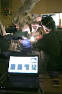
A biomedical engineer named Yoann Gosselin recently developed an imaging system that features a low light camera, a microscope, and a spectrometer and is used to detect a particular molecule that accumulates in malignant brain tissue, accurately revealing cancer cells.
MORE ARTICLES
Low light level cameras appear at Photonics West
Image sensor based on mantis shrimp vision detects cancer
Medical Imaging: Digital light projector localizes treatment of skin diseases
The imaging system is used as part of the surgical process of removing cancerous cells in the brain and when combined with radiotherapy and/or chemotherapy, will provide a better prognosis for brain cancer patients. In the system, Gosselin combined a Carl Zeiss neurosurgical microscope to a spectrometer, and coupled the latter to an HNü 512 EMCCD camera from Nüvü Cameras. The HNü 512 features a 512 x 512 EMCCD image sensor and achieves a frame rate of 67 fps. The camera, which is available with a PCIe Camera Link or GigE Vision interface, also has an EM gain range of 1-5000 and a spectral range of 250 – 1100nm.
With the addition of the HNü camera, the device detects the concentration of protoporphyrin IX (PpIX), a molecule that specifically accumulates in malignant brain tissue, with increased sensitivity. The camera helps by refining the boundaries between healthy cells for purposes of extracting cancer cells during surgery, while also providing a full image of the exposed brain area. In addition, the camera increases the amount of detectable diseased cells, which enhances the procedure’s efficiency and helps with prognosis and treatment.
The research was done in the radiology laboratory at l’École Polytechnique de Montréal and the system itself is set to be tested in the United States in clinical trial scenarios.
View more information onHNü cameras.
Share your vision-related news by contacting James Carroll, Senior Web Editor, Vision Systems Design
To receive news like this in your inbox, click here.
Join our LinkedIn group | Like us on Facebook | Follow us on Twitter | Check us out on Google +
About the Author

James Carroll
Former VSD Editor James Carroll joined the team 2013. Carroll covered machine vision and imaging from numerous angles, including application stories, industry news, market updates, and new products. In addition to writing and editing articles, Carroll managed the Innovators Awards program and webcasts.
