Stereo images speed glaucoma analysis
Andrew Wilson, Editor, [email protected]
Diseases of the optic nerve such as glaucoma cause a loss of retinal cells that results in the patient having a limited field of view. While untreated glaucoma may lead to optic nerve damage and blindness, the disease can often be detected early and treated with the correct medication.
“To diagnose diseases such as glaucoma,” says Bruno Laÿ, president of ADCIS (Hérouville Saint-Clair, France; www.adcis.net) and vice president of marketing & sales for Amerinex Applied Imaging (Monroe Twp., NJ, USA; www.AmerinexImaging.com), “a fundus camera is first used to produce photographic stereo pairs of each eye. By viewing these stereo pairs with specialized optical eyeglasses, the relief of the optical retina is highlighted and any possible disease rapidly diagnosed. However,” says Lay, “although digital fundus cameras are now available, many patient records are still archived on 35-mm photographic film.”
With recent US Food and Drug Administration (FDA) requirements mandating the use of digital images, Allergan (Irvine, CA, USA; www.allergan.com), a specialty pharmaceutical company that develops eye-care products, called upon ADCIS to develop a system capable of digitizing images from these 35-mm slides. As well, the system was required to capture patient information located on a 35-mm image frame and perform automatic diagnosis of any signs of glaucoma.
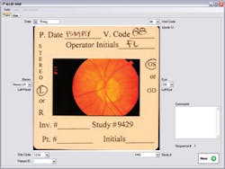
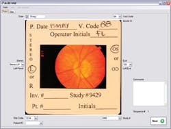
Allergan digitization system’s user interface shows a 35-mm slide fundus image, with notation fields on its frame.
As the system was required to digitize both the image of the retina and the patient information, a slide scanner could not be used. Instead, Laÿ and his colleagues developed a custom-built framing stand and mounted a Rebel XT digital camera from Canon (Lake Success, NY, USA; www.usa.canon.com) approximately 25 cm above the imaging platen. This was then interfaced to a PC running Windows XP over a USB 2.0 cable. Each image is digitized to 8 Mbytes per RGB channel resulting in a 24-Mbyte image file that is transferred to a RAID drive in the PC.
Once images are captured, the ADCIS software performs optical character recognition of the relevant data located in the frame. “Eight parameters around the frame of the image store the pertinent technical information about the image,” says Laÿ. “This includes the hospital and patient name, the visit code, whether the image is from the right or left eye, and which stereo pair image is present.”
Once these data have been determined the information can be displayed on the PC screen along with the two stereo image pairs of the eye. To register these two images for stereo viewing, they must first be properly aligned. To do this, the dark/light transitions at the edges of both images are computed and the center of each circle computed. The radius of each image can then be computed and the images aligned on the PC monitor for stereo viewing.
However, perhaps the most important feature of the system is its ability to discern whether any glaucoma is present. ADCIS, with the School of Mines (Paris, France; www.ensmp.fr), has developed sophisticated software to automatically process images of the retina and identify lesions. It is planned to use this software to automatically detect the optic nerve and to analyze images.
The software computes the brightest part of the image along a horizontal axis to determine the position of the optic nerve. Once these coordinates have been computed, a watershed transform is used to find the boundary of the optic nerve within the image. Three color histograms of the R, G, and B channels are then computed within this region of interest (ROI). “Interestingly,” says Laÿ, “any possible disease can be detected by examining the green histogram of this ROI, where if, for example, disease should be present, two peaks will appear.”
To make the software more widely available, ADCIS has commercialized many of the routines in a retinal analysis tool for the company’s Aphelion image-processing software. Although currently the tool is for research purposes only, the software can be integrated into ophthalmology systems depending on approval by agencies such as the FDA.

