Camera takes a closer look at the workings of the brain
Optical imaging of blood flow or oxygenation changes is useful for monitoring cortical activity in healthy subjects and individuals with epilepsy or those who have suffered a stroke.
However, to gain a better understanding of flow or oxygenation changes in conscious, freely moving animals, these optical systems must be implemented in a small-scale, portable design that can be worn by a patient.
Working with engineers fromQImaging, scientists at the University of Toronto are on their way to achieving that goal, having designed just such a prototype system for studying the effects of stroke recovery and epilepsy.
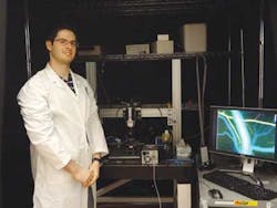
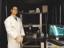
Using the system, University of Toronto assistant professor Ofer Levi has simultaneously measured blood oxygenation and changes in blood flow in the brains of anesthetized mice. The system also statistically distinguished between veins and arteries, something never before accomplished.
The system illuminates the brain tissue using multiwavelength vertical-cavity surface-emitting lasers (VCSELs) and captures images of the tissue using QImaging's Rolera 14-bit EM-C2EMCCD camera.
Using the setup, the researchers showed how flow and oxygenation changed in response to induced ischemia, a restriction in the blood supply to tissues that causes a shortage of oxygen and glucose needed for cellular metabolism.
Levi believes that the ability to obtain high spatial and temporal resolution with inexpensive equipment will made these optical imaging techniques a desirable alternative to commonly used clinical techniques such as fMRI, SPECT, or PET.
For a fully portable system, a smaller imaging device would be required, and Levi is building a system to enable optical neural imaging studies to be made of freely behaving animals, rather than ones that are constrained.

