3D Imaging: 3D tracking aids surgical navigation
During functional endoscopic sinus surgery, instruments such as endoscopes and surgical instruments are inserted through the nose to remove nasal polyps and treat sinusitis. Needless to say, avoiding entering the brain or injuring the eye are all paramount concerns during such surgical procedures.
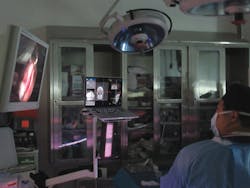
To overcome this, ClaroNav (Toronto, ON, Canada;www.claronav.com) has developed a system called NaviENT that dynamically determines the position of the tip of any surgical instrument in 3D using an optical triangulation technique. The position of the instrument is then mapped to a corresponding location in a pre-acquired CT scan of the patient's head allowing the surgeon to visualize the position of the instrument in the skull during surgery (Figure 5).
To locate the instrument's tip, the system employs a Bumblebee XB3 three-sensor multi-baseline IEEE-1394b stereo camera from Point Grey (Richmond, BC, Canada;www.ptgrey.com). The stereo camera features three 1.3Mpixel progressive scan CCDs, two of which are used for acquiring left and right monochrome images with a 1280 x 960 resolution at 16fps, while the third captures an overview of the scene. While the camera has two stereo baselines of 12cm and 24 cm, the extended 24cm baseline was chosen to provide higher precision at longer ranges.
In use, the XB3 camera on the system detects the location of black and white checkered patterns affixed to both the surgical instrument and the patient's forehead (Figure 6). Point Grey's FlyCapture SDK running under Windows 10 is then used to acquire the images. The position of the instrument relative to the patient tracker is calculated by ClaroNav's own proprietary optical tracking software to a fraction of a millimeter. "Our tracking device called MicronTracker detects the black and white pattern (called xpoints) and provides their location with accuracy of 0.2mm. EachXpoint provides 3D pose data and a combination of at least 3 XPoints are needed to represent a marker (or rigid body of 6DOF) in the space," says Ahmad Kolahi, CEO of ClaroNav Kolahi Inc.
During typical sinus and skull base procedures, surgery is performed under low light conditions. Hence it was important to create a system that is unobtrusive to the surgeon. To illuminate the scene, the NaviENT system uses an array of 2x96 IR LEDs of 5mm diameter, emitting at 850nm with a 60o field of view. These are mounted inside a tracking box with the Bumblebee XB3 camera. IR light projected onto the scene is then captured by the cameras and filtered with 850nm IR band-pass filters from Midwest Optical Systems (Palatine, IL, USA;www.midopt.com) prior to processing.
To overlay this information on the CT image, it is necessary to register the patient to the CT image. To do so, the surgeon marks landmark points on the CT image and identifies the corresponding landmarks on the patient. Having done so, the system automatically generates a registration map which correlates the two.
While the stereoscopic camera in the XB3 helps determineww the position of the instruments, the third center camera in the unit captures and displays live video. If the field of view of the stereoscopic camera becomes obstructed, the system is unable to detect the position of the instruments. If this occurs, a surgeon can refer to the live video feed to remove the obstruction prior to proceeding.
Unlike electromagnetic sensors, the proprietary optical tracking technology used in the NaviENT system does not require wired sensors and is not affected by the presence of metallic objects and electromagnetic fields in the environment. Furthermore, its accuracy does not degrade when targets are partly occluded or soiled.
The NaviENT system has been in clinical trials since 2013. Earlier this year, the NaviENT system received all technical certificates required for Health Canada and CE submission (such as 60601-1, EMC, 62471 photo-biological and biocompatibility) and in April 2016 the device was approved by Health Canada for sale in Canada.


