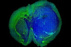
Laser imaging technique shows surgeons where tumors are in the brain
Using a technique called stimulated Raman scattering (SRS) microscopy, a team of researchers from Harvard University and the University of Michigan were able to build images which enabled them to distinguish between microscopic areas of tumor cells and healthy tissue in the brains of mice.
The technique was developed in 2008 by a team of Harvard researchers that included Xiaoliang Sunney Xie, the Mallinckrodt Professor of Chemistry and Chemical Biology and Minbiao Ji, a postdoctoral fellow in chemistry and chemical biology. It works by shining non-invasive lasers into tissue and detecting the weak signal that emerges. By analyzing the signal’s spectrum, researchers can build images of the cellular makeup of the tissue, according to the Harvard Gazette.
"Biopsy has been the gold standard for detecting and removing these types of tumors," said Xiaoliang Sunney Xie, the Mallinckrodt Professor of Chemistry and Chemical Biology. "But this technique, we believe, is better because it’s live. Surgeons can now skip all the steps of taking a biopsy, freezing, and staining the tissue. This technique allows them to do it all in vivo."
Working with a team of researchers led by Daniel Orringer, a physician and lecturer in the University of Michigan‘s Department of Neurosurgery, the Harvard team was able to demonstrate that their technique was effective in detecting tumors in living mice, and in human brain tissue, which was outlined in a September 4 paper in Science Translational Medicine.
In order to test the accuracy of the technique, the researchers performed a test in which they compared the method of Hematoxylin and eosin staining—the current approach used in brain tumor diagnosis—against SRS microscopy. In the test, three surgical pathologists trained in studying brain tissue and identifying tumor cells, had virtually the same level of accuracy regardless of which images were examined. The only difference being that SRS microscopy enables real-time analysis, according to Xie.
View the Harvard Gazette article.
View the research paper.
Also check out:
(Slideshow) Vision Systems Design 200th issue anniversary: Spanning the spectrum
Digital autopsies with 3D imaging software and medical scanners
How 3D visualization software could be used to help treat injured soldiers
Share your vision-related news by contacting James Carroll, Senior Web Editor, Vision Systems Design
To receive news like this in your inbox, click here.
Join our LinkedIn group | Like us on Facebook | Follow us on Twitter
About the Author

James Carroll
Former VSD Editor James Carroll joined the team 2013. Carroll covered machine vision and imaging from numerous angles, including application stories, industry news, market updates, and new products. In addition to writing and editing articles, Carroll managed the Innovators Awards program and webcasts.
