Imaging software tracks tumor rotation during radiotherapy
A method developed by researchers in Australia and Denmark uses intrafraction monitoring software to simultaneously estimate both the rotation and translation of a tumor during radiotherapy.
Previous studies have indicated that accounting for tumor rotation can facilitate a two to three millimeter reduction in the margins needed for treatment beams in prostate cancer patients, and reduced margins will lead to reductions in normal tissue toxicity and side effects, explained Paul Keall, senior author of the research paper and director of the Radiation Physics Laboratory at the University of Sydney in the Medical Physics Web article.
As a result, researchers from the University of Sydney and Aarhus University Hospital in Denmark decided to combine intrafraction imaging software technology and subsequent real-time treatment adaptation to produce what they predict to be the next technology to be adopted in cancer radiotherapy.
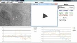
The technique works by estimating the 3D movement of markers implanted in the tumor from 2D kilovoltage images acquired continuously during treatment by a kV x-ray imager mounted perpendicularly to the radiation treatment beam source. The markers in the tumor are automatically segmented by imaging software and the rotation around three axes—right–left, superior–inferior, and anterior–posterior—is estimated by using the iterative closest point algorithm. This algorithm calculates the rotation needed to transform the marker coordinates at a given time to the original source coordinates detected in images acquired prior to treatment. It was found to converge to an optimum transform in two to three iterations, with the translation and rotation calculated in less than 300 milliseconds after the initial target motion, according to the article.
In tests using in vivo images from 10 cancer patients, as well as simulated images, the algorithm was applied effectively with just a sub-millimeter error rate. (For the simulated images, the root mean square error rate, or the difference between the predetermined location and those derived from the images, was an average of 0.004°. For the in vivo images, the algorithm was applied successfully, showing the measured tumor motion to be in line with other prostate studies. Full clinical testing of the algorithm and study are expected to occur at some point during 2014.
View the Medical Physics Web article.
Also check out:
Chemical imaging technique could improve diseased tissue analysis
Optical sensor tracks zinc in cells for cancer research
Fraunhofer imaging technique enables faster testing of drug ingredients
Share your vision-related news by contacting James Carroll, Senior Web Editor, Vision Systems Design
To receive news like this in your inbox, click here.
Join our LinkedIn group | Like us on Facebook | Follow us on Twitter | Check us out on Google +
About the Author

James Carroll
Former VSD Editor James Carroll joined the team 2013. Carroll covered machine vision and imaging from numerous angles, including application stories, industry news, market updates, and new products. In addition to writing and editing articles, Carroll managed the Innovators Awards program and webcasts.
