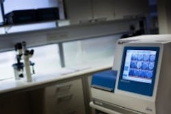
Embryoscope imaging device may improve chances of In Vitro Fertilization
A device called the Embryoscope combines an incubator that maintains ideal temperature and air conditions with an imaging system that provides time-lapse video of an embryo’s development, which one physician is calling the most exciting breakthrough since In Vitro Fertilization began.
Developed by FertilTech—a company whose sole aim is to create technology to improve human embryo assessment in assisted reproduction— the Embryoscope features an imaging system that consists of a Leica 20 x 0.40 LWD Hoffman contrast objective microscope specialized for 635 nm illumination. The microscope features a 1280 x 1024 pixel focal plane array with 3 pixels per µm and 8-bit digital output and utilizes a single 1W red LED light (635 nm). With the Embryoscope, physicians can acquire images of the embryo at a rate of 10 minutes per six slides (60 embryos).
The Embryoscope also features a tri-gas incubator that maintains a temperature range of 86°F to 113°F and circulates and regenerates the gas volume of the incubator every 10 minutes. An embedded system based on an Intel Core duo T2300E 1.66 GHz processor controls the incubator. This system features Ethernet and USB ports for data exchange and a 12.1” embedded touch screen for display purposes.
Embryoscope devices capture images every 10-15 minutes, creating a time lapse video of the embryo’s development, which eliminates the need to expose the embryo to the outside atmosphere, and provides embryologists with additional information with which to make decisions.
"I think this is the most exciting breakthrough since IVF started," Dr. Simon Fishel, managing director of the UK's CARE Fertility, told CNN.
He added, "The information that we are gathering with the Embryoscope with the time-lapse is far superior," he says. "We have much, much more information on which to base the crucial decision as to which embryo is the one to transfer back to the patient."
Fishel was responsible for the first Embryoscope baby born in the UK, in 2012, as well as the first ever IVF baby back in 1978. CARE estimates that its use of the Embryoscope and learning algorithms has increased the chance of pregnancy by about 20%.
The article explains that embryos with early abnormalities can be immediately ruled out, and learning algorithms have been created to positive or negative patterns at key development points thereafter –meaning patients can be implanted with embryos that have an optimal chance of success.
Use of the Embryoscope has increased in IVF facilities around the world, but barriers remain, according to CNN. First, the vast amount of extra data requires greater resources for analysis, and second, the technology itself is expensive.
View more information on the Embryoscope.
Also check out:
Medical Imaging: Polarization subtraction system characterizes cancer
Researchers developing terahertz detectors with eye on improving certain imaging applications
Machine vision content collections: USB 3.0, medical imaging, and smart cameras
Share your vision-related news by contacting James Carroll, Senior Web Editor, Vision Systems Design
To receive news like this in your inbox, click here.
Join our LinkedIn group | Like us on Facebook | Follow us on Twitter | Check us out on Google +
About the Author

James Carroll
Former VSD Editor James Carroll joined the team 2013. Carroll covered machine vision and imaging from numerous angles, including application stories, industry news, market updates, and new products. In addition to writing and editing articles, Carroll managed the Innovators Awards program and webcasts.
