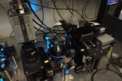
Scientific CMOS cameras produce images of embryos in real time
Researchers from the Max Planck Institute and the Technical University in Dresden, Germany have created a microscope that uses two Andor Zyla scientific CMOS cameras to capture and produce images of an embryo in real time.
Andor Zyla 5.5 MPixel (2561 x 2160 pixel) cameras feature a frame rate of 100 fps (Camera Link 10-tap) and 30 fps (Camera Link 3-tap), front-illuminated scientific CMOS image sensor, Camera Link interface, and global and rolling shutter. The microscope used the two Zyla cameras in a specially-designed four-lens selective plane illumination microscope with on-board image processing. With this microscope, researchers were able to produce undistorted, high-resolution images of the entire endoderms of multiple zebrafish embryos in less than ten seconds, according to a press release.
Jan Huisken, research group leader at the Max Planck Institute, said that the spherical geometry of the embryos was exploited, and that the speed and sensitivity of the Zyla cameras enabled them to compute a radial maximum intensity projection of each individual embryo during image acquisition.
"An entire zebrafish embryo can now be instantaneously projected onto a 'world map' to visualize all endodermal cells and follow their fate. This reveals characteristic migration patterns and global tissue remodeling in the early endoderm and, by merging data from many samples, we have uncovered stereotypical patterns that are fundamental to embryo development."
In addition, Huisken said that while this technique will not replace 3D imaging techniques, it should streamline future research endeavors.
"The raw data from the Zyla cameras were not saved at any point, which means that any 3D information not captured in the projection is lost," he said. "However, the radial projection delivers immediate, pre-processed data for analysis and enables experiments to be repeated very rapidly. This new technique will not eliminate the need for slow 3D imaging techniques in more complex shapes but it is a highly effective strategy to streamline further analysis and increase throughput in many applications."
View more information on Andor sCMOS cameras.
Also check out:
MIT and NASA researchers develop microscope prototype that uses neutrons
DARPA spends $10 million for stronger, stealthier vision-guided military robot
Scottish researchers using 3D imaging to view tongues in speech study
Share your vision-related news by contacting James Carroll, Senior Web Editor, Vision Systems Design
To receive news like this in your inbox, click here.
Join our LinkedIn group | Like us on Facebook | Follow us on Twitter | Check us out on Google +

James Carroll
Former VSD Editor James Carroll joined the team 2013. Carroll covered machine vision and imaging from numerous angles, including application stories, industry news, market updates, and new products. In addition to writing and editing articles, Carroll managed the Innovators Awards program and webcasts.
