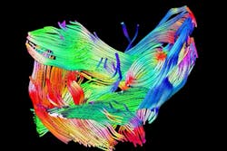
MRI system images muscles in 3-D
Researchers at the Technische Universiteit Eindhoven (TU/e; Eindhoven, The Netherlands) and the Academic Medical Center (AMC) in Amsterdam have developed a technique that allows muscle structures to be imaged in 3-D.
The technique uses what is known as MRI diffusion tensor imaging which allows the movements of water molecules in living tissue to be viewed.
This was already possible on a small scale with simple muscles, but thanks to Eindhoven University's Dr. Martijn Froeling, who improved upon the speed at which MRI data could be acquired, it can now also be performed on a larger scale and with complex muscle structures. More importantly, the improved technique also reveals very small muscle damage.
To study the effectiveness of the technique, Froeling imaged a range of subjects including the thighs of marathon runners at different times: one week before a marathon, two days after it, and again three weeks after. He was able to visualize the muscle damage following the marathon, which was still visible after three weeks, even though many of the runners no longer reported any pain in their muscles.
AMC Amsterdam and TU/e now intend to use the technique in studies of post polio syndrome and spinal muscular atrophy.
More information on the system is available here.
Recent articles on medical imaging that you might also find of interest.
1. Medical imaging system comes above the radar
Bristol University spin-out Micrima (Bristol, UK) is developing a medical imaging system that can distinguish a breast tumor from normal tissue by detecting the difference between their dielectric properties.
2. Wafer-scale imager targets medical imaging applications
Engineers at the Science and Technology Facilities Council’s (STFC; Didcot, UK) Rutherford Appleton Laboratory (RAL) have designed a wafer-scale digital image sensor for mammography and digital tomosynthesis applications.
3. Researchers turn an iPhone into an otoscope
A pediatric medical device being developed at Georgia Tech (Atlanta, GA, USA) and Emory University (Atlanta, GA, USA) could make life easier for parents with children suffering from an ear infection.
4. Imaging system gets under the skin
European researchers have used optical coherence tomography (OCT) to noninvasively image the network of blood vessels beneath the outer layer of skin, potentially revealing telltale signs of disease.
5. Imaging combination delivers high-contrast, high-resolution images
Scientists from the University of Southern California (Los Angeles, CA, USA) and Washington University (St. Louis, MO, USA) have developed a new type of medical imaging system that gives doctors a new look at live internal organs.
Vision Systems Design magazine and e-newsletter subscriptions are free to qualified professionals. To subscribe, please complete the form here.
-- Dave Wilson, Senior Editor, Vision Systems Design
