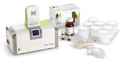
Camera performs colony counting
To monitor thequality of pharmaceutical ingredients, it is critical to test for microbial contamination throughout the manufacturing process. Detecting the amount of bacterial contamination present in a sample is achieved by passing the sample through a filter of a particular pore size. The micro-organisms remain on the filter surface; when the filter is placed in a Petri dish and saturated with an appropriate medium, the microbes grow into colonies, which can then be counted.
Such traditional microbiological methods are slow, due to the lengthy incubation times required to grow the colonies, and results can take several days to obtain. Seeking to reduce the incubation time, engineers atMillipore (Billerica, MA, USA) have developed a system called the Milliflex Quantum that combines industry-standard membrane filtration techniques with fluorescent staining, making the colonies visible to the naked eye in a much shorter period of time.
Not content with having done so, the engineers also decided to incorporate a system into the unit that could be used to capture images of the colonies, after which they could be displayed on a PC. To help them with the task, they turned to Stemmer Imaging (Puchheim, Germany).
The Stemmer engineers created the image-acquisition hardware around auEye board-level camera from IDS Imaging (Obersulm, Germany) with a monochrome 1280 × 1024-pixel resolution CMOS sensor and a 12-mm lens. GPVision (Chateaneuf Sur Loire, France), one of Stemmer's integration partners, then developed the supporting user interface and control software.
The camera module—which was originally developed as an option for the Quantum system—has already been tested by a number of customers, 80% of whom said that they preferred to use the system with the camera rather than without it. As a result, the camera is now a standard feature of the system.
More Vision Systems Issue Articles
Vision Systems Articles Archives
