Filters open new vision applications
Specialized light sources and filters open imaging options in industries ranging from biomedical and pharmaceutical to metal finishing and lumber.
By R. Winn Hardin, Contributing Editor
The art of lighting in machine vision is often referred to as a “black art”-black meaning the absence of light. However, machine vision starts with light, and its success depends on it. If the light is the wrong type, installed incorrectly, housed inefficiently, or mismanaged in some other way, then the machine-vision application is doomed to failure.
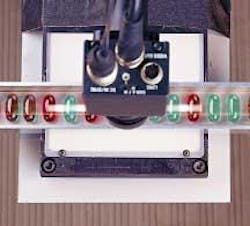
FIGURE 1. Combining high- and low-bandpass filters with broadband light sources can improve machine-vision color inspections by enhancing contrast, even when the system uses a monochrome camera for pharmaceutical applications such as inspecting pills in blister pack.
Machine-vision specialists use a variety of filters and enclosures with solid-state, fluorescent, or gas-discharge lamps, as well as monochromatic lasers, to assist with machine-vision applications. This development has led to increased understanding of illumination sources and filters in the vision industry, which, when combined with improvements in sensors and image processing, is driving machine vision into new application areas such as drug discovery and medicine (see Fig. 1). Existing markets are benefiting, too, with specialized light sources enabling new industrial applications, such as blind lot codes, ambient light, and spurious reflection filtering.
STRONG CONTENDERS
Prior Scientific sells material-handling equipment and light sources to the semiconductor, industrial, and biotechnology markets, according to sales manager Dennis Doherty. “We are seeing more requests for wavelength-specific or filtered illumination from biological and medical applications, such as where a customer is doing fluorescence imaging to see what a protein attached to a cell does,” Doherty says. “Most industrial or semiconductor work that we’re involved in uses brightfield imaging without a lot of filters.”
For example, one of the most popular fluorescent tags, green fluorescent protein (GFP), latches onto cellular structures and then absorbs light at 400 nm with a secondary peak-absorption line at 475 nm (blue). The GFP converts those photons to electrons and then re-emits photons or fluoresces at 509 nm. Biologists track this fluorescence to see how chemicals and other operations affect a cell as the protein moves through it. According to Doherty, OEMs selling automated testing equipment into the pharmaceutical and biotechnology markets typically use a high-resolution CCD camera with a bandpass filter external to the C-mount lens assembly that blocks all incoming light except light near the GFP’s emission peak around 509 nm.
Manual experiments may use a laser that emits at or near 400 nm; however, automated systems prefer a broadband light source for two reasons, Doherty says. Lasers are expensive and sometimes difficult to use, and they are spectrum-limited. Lasers are designed to emit in a narrow spectral band that limits how many different fluorescent markers (and, therefore, experiments) a system can conduct without requiring a different laser.
Intense lamp sources, such as metal halide, xenon, or mercury gas-discharge lamps, offer broadband illumination that can be filtered to specific wavelengths and still provide enough photons to generate a detectable fluorescent signal. Because of the size, heat, and electrical-power requirements for these lamps, integrators typically use a fiberoptic liquid lightguide to deliver the light to the sample, Doherty says. These fibers have internal reflection coating on the inside of the hollow optical fiber and liquid inside. Nonliquid fiberoptic bundles have small gaps between the individual fused fibers, which results in light loss. These fibers also are smaller than the 3-mm apertures afforded by liquid lightguides, making it more difficult to integrate the lightguide, lamp, and collecting optics in the illumination system, compared to liquid fibers.
“For every photon that hits a sample, you want to collect the subsequent fluorescence emission. The more light you have, the more emission you get. A light source emitting at 100% that goes through a neutral-density filter and/or bandpass filters and optics may only deliver a few percent of the initial power. So you really need intense lights, which is why they use the [high-intensity] lamps,” says Doherty. “However, if the application calls for a light source below 350 nm [in the ultraviolet (UV)], above 650, or 680 nm [in the infrared (IR)], you have to go to specialty light sources.”
Matching the absorption spectrum from the most likely fluorescent markers to the lamp’s output spectrum is considered the best way to match a light source with a biological application, he adds. For instance, mercury is strong in the blue, which works well with GFP, while metal halide is relatively flat through the visible spectrum, making it attractive for systems that use a variety of markers. Another way to classify peak output for a lamp is through color temperature.
The table above indicates typical color temperatures for a variety of sources. As the temperature increases, the peak moves toward the blue and UV part of the spectrum; as it decreases, the peak moves toward the red and IR parts of the spectrum.
MONOCHROME CAMERAS
Combining high- and low-bandpass filters with broadband light sources also can improve machine-vision color inspections by enhancing contrast, even when the system uses a monochrome camera. According to Jeff Harvey, marketing manager for Edmund Industrial Optics, pharmaceutical applications such as inspecting pills in blister packs often use this approach.
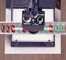
“During packaging, pharmaceutical pills of different colors need to be sorted,” says Harvey. “Pills are inspected for specific characteristics as they travel down a trough-like conveyor belt prior to sorting. A minimum of 60% contrast is needed for the software to differentiate between the different pills.” Working at a distance of 350-450 mm and a required field of view of approximately 70 mm, Edmund Optics suggests a 35-mm MVO double-Gauss imaging lens with 1/2-in. CCD camera, such as the Sony XC-75 monochrome camera. An unfiltered fiberoptic area backlight illuminates the red and green pills; a bandpass filter is placed on the camera’s optics; and a Coreco Imaging Bandit frame grabber captures the camera signal for processing (see Fig. 2).
Figure 2. To differentiate between colored pills, software needs a minimum of 60% contrast. A gray-scale profile can be generated from the sample area to calculate the contrast. The original monochrome image has only 8.7% contrast difference between red and green pills. The contrast can be increased beyond the minimum requirement by ~25% by attaching a green filter to the front of the lens. This allows the user’s customized software to operate on a go/no-go principle and accurately sort the pills.
“Monochrome cameras cannot inherently discriminate between different colors,” explains Harvey. “Red and green pills appear nearly identical when imaged with the Sony XC-75 camera. Filtering can be used to improve the contrast between pills of different colors and enables the system to differentiate between them.” Edmund uses the equation
% contrast = Imax − Imin/Imax + Imin × 100
No filter: Contrast = 119 − 100/119 + 100 = 8.7%
Red filter: Contrast = 217 − 62/ 217 + 62 = 55.6%
Green filter: Contrast = 166 − 12/166 + 12 = 86.5%
The green filter exceeds the contrast requirements dictated by the image-processing algorithms; therefore, the green filter was installed in this application.
LEDS AND LASERS
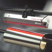
Light-emitting diodes (LEDs) typically eliminate the need for a filter at the light source. LEDs also offer longer lifetimes than most lamps, are more efficient at electrical-to-light conversion, and are smaller and easier to direct because of their small aperture. With typical outputs per diode in the milliwatt range, however, LEDs are generally less-intense illumination sources than high-intensity lamps or lasers. If LEDs meet the application’s intensity needs, integrators can use an LED’s color specificity and other operational benefits to enhance a vision application (see Fig. 3). “If your application requires intense illumination, then a high-intensity quartz halogen or tungsten halogen lamp source is generally the lighting source of choice. LEDs work fine as long as there’s enough intensity,” adds Joe DiRuzza, StockerYale director of sales and product management.
FIGURE 3. StockerYale Cobra 500 LED line illuminator provides working intensities up to 500,000 lux of 630-nm (red) light and is especially suitable for web-surface-inspection applications.
Daryl Martin, Midwest sales and support manager for Advanced Illumination, says one of the most common uses of red LEDs and a matching bandpass filter is to greatly reduce the effect of ambient lighting on vision in the automotive industry. “It’s easy to get strong reflections from fluorescent lights on the ceiling in an automotive manufacturing plant,” Martin explains. “To avoid that, you use a red LED with matched bandpass filter. Fluorescent lights do not have strong red content, so the result is a reduction in negative content from ambient lighting of 35 times. If you’re trying to block sodium, which does have strong red content, then the effectiveness of a red bandpass filter for blocking ambient light is diminished.”
According to StockerYale’s DiRuzza, LEDs work best when the wavelength of the LED complements the object under inspection. For instance, green LEDs work well on copper surfaces because copper reflects red light and absorbs green light, which highlights surface defects. Red light does the same for black surfaces, while the new white-light LEDs work for optical-character-recognition applications. “The blue/white LED light on black-and-white printing raises the print, giving the black type more definition,” DiRuzza says. New UV LEDs are making inroads in blind-lot reading for pharmaceutical and food containers using UV-sensitive inks. The ink absorbs UV light and emits in the visible so that standard machine-vision cameras can read the “blind” lot code.
StockerYale Canada regional sales manager Sylvain Bosse suggests that customers consider laser-line generators rather than broadband light sources for applications that require multiple light sources of different colors. “For example, one of our customers uses a few cameras, filters, and lasers with different wavelengths for lumber inspection,” Bosse said. “Using multiple line generator lasers, a line is projected around the full circumference of the log to measure its size and find defects by triangulation. Each camera looks at a section of the line (at a specific laser) and is equipped with a filter to avoid capturing the lines projected by the other lasers.
Another customer is using the same principle to inspect a cylinder-shaped section of an anode used in aluminum foundries. “Laser line scanners are good at generating 3-D coordinates in applications where the laser is stationary and the object moves through the laser inspection station at high speeds. Lasers provide plenty of light for cameras working at high rates. Systems that use broadband sources projected through gratings for 3-D measurements deliver less light per unit area compared with a laser and, therefore, can be limited by available light and the resulting contrast at the camera/sensor.
Whether it’s a single-wavelength laser, narrow-bandwidth LED output, or broadband fluorescent lamp, integrators have many illumination choices. This will continue as manufacturers use more UV-sensitive dyes and tracers and color packaging becomes the norm.


