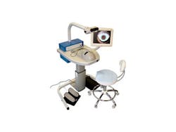
Scope system simulates eye surgery
Engineers at VRmagic (Mannheim, Germany) have developed a simulator that can help prospective eye surgeons train for operations.
The Eyesi eye operation system consists of a microscope through which surgeons can view simulated operations while guiding medical instruments into a mechanical eye situated in a model of the head.
Both the eye and the instruments are color coded to enable the simulator to determine their position and alignment. To do so, a field-programmable gate array (FPGA)-based camera captures images of the instruments and the eye and transmits their position via a USB cable to a computer. There, the 2-D coordinates for every color marker are determined, and from this data a 3-D image is created.
Based on the 3-D data, biomechanical simulation algorithms developed for the system can then calculate in real time the way in which the eye should behave when it interacts with the instruments.
The simulated image of the interior of the eye is displayed for both the left and the right eye on an OLED display in the simulator’s stereo microscope.
-- By Dave Wilson, Vision Systems Design
