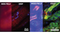
Computational and NIR imaging increase wide-field microscopy depth and resolution at increased speed
Researchers from MIT (Cambridge, MA, USA: www.mit.edu) and Harvard University (Cambridge, MA, USA; www.harvard.edu) developed an imaging technique called DEEP (De-scattering with Excitation Patterning) that uses near-infrared light and computational imaging algorithms to improve over existing point-scanning techniques. This improvement could allow researchers in the future to capture high-definition images of blood vessels, or even individual neurons in the brain, at previously unattainable speeds.
Point-scanning multiphoton microscopy shines near-infrared or shortwave infrared light onto a point to image. Fluorescent molecules at that point absorbs two or three photons of laser light and emits a photon in visible wavelength; one pixel in the image corresponds the number of emitted photons from that point. Point-scanning creates high quality images, but the process is slow.
Wide-field multiphoton microscopy, a faster technique, images an entire plane in a tissue sample versus a single point, at the cost of degraded resolution. The wide-field method also is only effective at shallower depths compared to the point-scanning method due to the scattering of light as it reflects off the plane.
In their paper titled “De-scattering with Excitation Patterning enables rapid wide-field imaging through scattering media,” the researchers describe a method of removing the background noise that occurs when employing wide-field imaging methods, to provide the same resolution provided by point-scanning methods.
The deeper the plane a wide-field technique images, the more high-frequency information degrades. Low frequency information is generally retained regardless of the depth of the scan. By projecting a wide-field pattern of light versus even illumination onto a sample to image, individual points can still be imaged even in wide-field image.
Computational imaging then combines the data recorded for each individual point into a single image. The final image mimics the product of a wide-field scan while retaining the resolution of point-scanning. This “best of both worlds” approach allowed the researchers to double the previous possible depth of wide-field two-photon imaging.
“We have demonstrated 100-1000 times speed improvement over the state of the art in two-photon computational imaging techniques,” says Dr. Dushan Wadduwage, one of the researchers. “But we haven’t experimentally demonstrated the same over point scanning techniques. This is because point scanning microscopes have been around for decades and are fully optimized in every way. Our experimental design should still be optimized in terms of the sensor and power.”
“But in theory if we consider all these aspects the same for point-scanning and DEEP, we will be 100-1000x faster than the point scanning two-photon microscopes,” says Wadduwage.
Potential, practical uses of the new technique include rapidly imaging blood vessels in mice to study how neurodegenerative diseases like Alzheimer’s affect blood flow or studying individual neurons or tumors by combining this method with use of fluorescent dyes or calcium probes.
About the Author

Dennis Scimeca
Dennis Scimeca is a veteran technology journalist with expertise in interactive entertainment and virtual reality. At Vision Systems Design, Dennis covered machine vision and image processing with an eye toward leading-edge technologies and practical applications for making a better world. Currently, he is the senior editor for technology at IndustryWeek, a partner publication to Vision Systems Design.
