Imaging technique could detect blood clots in a single scan
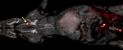
Seen on ScienceDaily:A blood clot can potentially trigger heart attacks, strokes and other medical emergencies. Treatment requires finding its exact location, but current techniques can only look at one part of the body at once. Now, researchers are reporting a method, tested in rats that may someday allow physicians to quickly scan the entire body for a blood clot.
Read full article onScienceDaily.
Our take:
By taking an innovative twist on traditional positron emission tomography (PET) imaging, researchers from Massachusetts General Hospital believe they have developed amedical imaging method that will enable healthcare providers to perform a single scan of a body for a blood clot.
In traditional methods of detecting a blood clot, a physician may need to use three methods: ultrasound to check the carotid arteries or legs, magnetic resonance imaging (MRI) to scan the heart and computed tomography to view the lungs.
"It's a shot in the dark," said Peter Caravan, Ph.D "Patients could end up being scanned multiple times by multiple techniques in order to locate a clot. We sought a method that could detect blood clots anywhere in the body with a single whole-body scan."
The approach, which is being presented at the 250th National Meeting & Exposition of the American Chemical Society, was developed by Caravan and his team at the Martinos Center for Biomedical Imaging at Mass. General. In the new method, the researchers developed a blood clot probe by attaching a radionuclide to a peptide that binds specifically to fibrin, which is a protein found in blood clots. Radionuclides can be detected anywhere in the body through PET imaging, so the researchers used different radionuclides and peptides, as well as different chemical groups, for linking the linking the radionuclide to the peptide, to identify which combination would provide the brightest PET signal in blood clots.
In order to test it out, the researchers studied how well the probe detected blood clots in rats. After testing 15 candidate blood clot probes, the team identified the best-performing probe, called FBP8, which contained copper-64 as the radionuclide.
The big question from here, according to Caravan, is how well these will perform in patients. The team is hoping to start testing the probe in human patients in the fall, but it could take five years of additional research before it is approved for use in a clinical setting.
While still in the testing phases with much work ahead, the progress made is positive and could lead to a streamlined process for blood clot testing, which is a benefit to both physician and patient.
- James Carroll, Senior Web Editor
You might also like:
