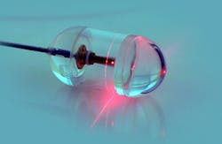
Capsule captures images of the esophageal wall
Researchers at the Wellman Center for Photomedicine at Massachusetts General Hospital (MGH; Boston, MA, USA) have developed an imaging system enclosed in a capsule about the size of a multivitamin pill that creates detailed, microscopic images of the esophageal wall.
The system may provide a new way to screen patients for Barrett's esophagus -- a precancerous condition usually caused by chronic exposure to stomach acid -- that doesn't require patient sedation, a specialized setting and equipment, or a physician who has been trained in endoscopy.
"By showing the 3-D, microscopic structure of the esophageal lining, it reveals much more detail than can be seen with even high-resolution endoscopy," says Professor Gary Tearney of the Wellman Center and the Pathology Department at MGH.
The capsule developed by Tearney and his colleagues houses a rapidly rotating laser that emits a beam of near-infrared light and sensors that record light reflected back from the esophageal lining. The capsule is attached to a string-like tether that connects to the imaging console and allows a physician or other health professional to control the system.
After the capsule is swallowed by a patient, it is carried down the esophagus by normal contraction of the surrounding muscles. When the capsule reaches the entrance to the stomach, it can be pulled back up by the tether. Images are taken throughout the capsule’s transit down and up the esophagus.
"The images produced have been some of the best we have seen of the esophagus," says Tearney. "We originally were concerned that we might miss a lot of data because of the small size of the capsule, but we were surprised to find that, once the pill has been swallowed, it is firmly 'grasped' by the esophagus, allowing complete microscopic imaging of the entire wall."
Related items on medical imaging that you might also be interested in reading.
1. Infrared cameras enhance diagnostic medical imaging
Many machine-vision applications rely on visible-light cameras; however, more specialized medical applications demand imaging in the nonvisible spectrum.
2. MRI system images muscles in 3-D
Researchers at the Technische Universiteit Eindhoven (TU/e; Eindhoven, The Netherlands) and the Academic Medical Center (AMC) in Amsterdam have developed a technique that allows muscle structures to be imaged in 3-D.
3. Hand held instrument inspects the retina
Medical technology firm Lumetrics (Rochester, NY, USA) has won a $973,000 grant from the National Institutes of Health (Bethseda, MD, USA) to fund the development of a digital hand-held diagnostic ophthalmic instrument for inspecting the human retina.
-- Dave Wilson, Senior Editor, Vision Systems Design
