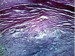
Artery walls imaged in vivo
Patients undergoing angioplasty or other heart-related medical procedures could benefit from a new technology being developed with funding from the National Science Foundation (NSF).
Optical imaging technology from Wasatch Photonics (Logan, UT, USA) creates images of coronary artery walls in vivo to show where lesions and plaques have formed. Physicians can use the images to determine the best course of action, including where a stent might be placed.
William J. Brown, vice president of business development at Wasatch Photonics, said the outcome of developing the technology will be the availability of a new tool to identify and treat coronary artery disease.
"This disease affects an estimated 16m Americans and is a primary cause of heart attacks and strokes. Identifying and treating plaque buildup and other intravascular conditions could reduce the morbidity and mortality rates from coronary artery disease," he says.
Wasatch Photonics technology, called intravascular optical coherence tomography, provides detailed imaging information for physicians to use in plaque assessment and stent implantation and monitoring.
Wasatch Photonics received a two-year Small Business Innovation Research (SBIR) Phase II grant for $498,325 from the NSF. The grant provides funding to continue developing the intravascular optical coherence tomography system.
Related items from Vision Systems Design that you might also find of interest.
1. Silicon Valley imaging outfit forges academic partnership in Ireland
Compact Imaging (Mountain View, CA, USA) has formed a two-year research collaboration with academics at the National University of Ireland Galway (NUI; Galway, Ireland) and University of Limerick (Limerick, Ireland) to further develop the company's optical coherence tomography (OCT) technology.
2. Imaging system gets under the skin
Researchers from Medical University Vienna (MUW; Vienna, Austria) and the Ludwig-Maximilians University (Munich, Germany) have used optical coherence tomography (OCT) to noninvasively image the network of blood vessels beneath the outer layer of skin, potentially revealing telltale signs of disease.
3. 3-D imaging goes under the microscope
A team of engineers at Swiss-based Heliotis (Root Längenbold, Switzerland) have developed a novel 3-D sensor based the principle of parallel optical low-coherence tomography (pOCT).
-- Dave Wilson, Senior Editor, Vision Systems Design
