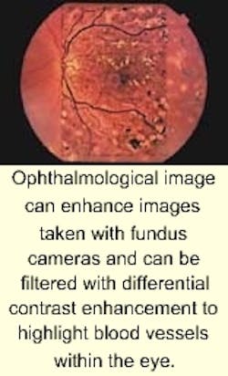
Imaging software speeds eye procedures
Certain eye disorders, such as diabetic retinopathy, affect the retinal circulation. Tests using fluorescein dye and fundus cameras can photograph the structures in the back of the eye and are useful in finding leakage or damage to the blood vessels that nourish the retina.
In tests, a colored dye is injected into a vein in the arm of the patient. The dye travels through the circulatory system and reaches the vessels in the retina and those of a deeper tissue layer called the choroid. After the dye has passed through the blood vessels to the eye, any vascular lesions become visible.
To diagnose eye-related diseases with certainty, the dye's departure from the eye's blood vessels must be documented in a series of images. Today's fundus cameras such as the PC-controlled FF-450 from Carl Zeiss Optical (Chester, VA) can simultaneously accommodate two 35-mm cameras and a video CCD camera or one 35-mm camera and up to three video or digital cameras.
For fluorescence angiography to be performed properly, flash lighting must be used to obtain the fluorescence needed from the fluorescein dye. Using analySIS software from Soft Imaging System (Lakewood, CO) in conjunction with a flash-control interface from PSI Pawlowski Medizintechnik (Jena, Germany), cameras such as the FF-450 can accelerate many aspects of eye examination.
To automate this procedure, both analog and digital images from the fundus camera are transferred to the system's host PC using a Soft Imaging Systems GrabBit frame grabber. After images appear on-screen, they can be archived along with patient data. AnalySIS software controls the acquisition sequence, beginning with the swiveling into position of the excitation filter, flash ignition, image acquisition and display. As soon as the flash is charged, the next acquisition is made.
Images in a series are given the same name, supplemented by a sequential number. Each image can be saved in the database immediately after acquisition or upon completion of the whole exam. This is advantageous if the user wishes to insert only select images into the database, or if image optimization via focus or differential contrast enhancement filters are necessary.

