ASIC-based imaging enhances MRI analysis
Image enhancement—the suppression of "noise" in digital images—is critical for modern medical technologies such as magnetic resonance imaging (MRI), computed tomography, and digital x-ray. To preserve the desired image information during enhancement without modification is a technical challenge, because most methods used in automated image analysis and image enhancement are apt to change the details or structures in the image.
ContextVision (CV; Great Falls, VA) was approached by health-care provider organizations that required increased performance at lower cost in the re-engineering of its MRI accelerator board. Unable to interest commercial IC vendors to build a specialized component, CV decided to build a custom ASIC.
"ASIC design was outside our competency," says Hans Norberg, CV's project manager for hardware development, "but we did produce detailed specifications on exactly what we needed and how it had to work." The company then commissioned Synopsys (Mountain View, CA) to produce the ASIC using CV's technology at speeds exceeding those of former accelerator boards.
Conventional image processing generally assumes that each individual pixel has a particular significance, for example, the intensity or color information about a pixel determines its relevance to the image as a whole. In many applications, this approach is sufficient. However, a more accurate method is required in more-complex image structures, where the significance of individual pixels can only be realized in a contextual environment. In most images, data levels create a hierarchical structure, with the lowest level representing the original image and higher levels representing more detailed information about the image, that is, edges and lines, their orientation and curvature.
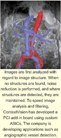
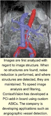
In most image-processing tasks, a one-level analysis defines the final result. However, in CV's image-enhancement method, images from higher levels control the operations performed on lower levels. When no structures are found, noise reduction is performed, and where structures are detected, they are maintained. This method can detect and retain information and sharpen and clarify structures.
ContextVision's algorithms evaluate whether every pixel in the image is part of the structure or part of the visual noise surrounding the structure. Image-enhancement based on adaptive filtering techniques strengthens the image and reduces the noise. Filtering increases the image's signal-to-noise ratio (S/N) by running algorithms that heavily filter noisy areas and provide sharpening in high S/N areas.
To embed these algorithms in hardware, Synopsys' designers needed to produce an ASIC so that the four-to-eight devices could be mounted on a single PCI-bus card. The resulting chip, named Javelin, designed with 0.35-µm technology, contains almost a half-million gates with embedded-memory functions and performs the equivalent of thirty-two 16 x 16 multiplications when running at 40 MHz.
The first accelerator board, having a four-chip configuration, will cut image-enhancement processing time to seconds for small images. Based on the design, CV has developed a PC-based system, called SharpView, that either provides image quality comparable to newer MRI systems at higher field strengths or maintains image quality at considerably shorter scanning times (see image on p. 8).
SharpView is a stand-alone, PC-based image-enhancement workstation used for MRI applications that consists of image-enhancement software and an accelerator board installed in a PC connected to an MRI scanner. In clinical trials, research proved that an operator using a low-field MRI scanner took approximately 45 minutes to scan each patient. After installing SharpView, the average scan time was decreased by 30%.

