Spectroscopy aids in characterizing skin cancers
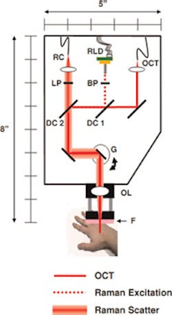
A joint US-Dutch team under the direction of Anita Mahadevan-Jansen from Vanderbilt University (Nashville, TN, USA) has developed a noninvasive probe capable of both morphological and biochemical characterization of skin cancers.
The portable instrument combines a Raman spectroscopy (RS) system with an optical coherence tomography (OCT) device and enables the sequential acquisition of co-registered OCT and RS data sets. An Andor (Belfast, Northern Ireland) high-resolution, near-infrared-enhanced Newton camera was chosen as the core element of the Raman diagnosis module.
The probe screens large areas of skin up to 15 mm wide to a depth of 2.4 mm with OCT to visualize microstructural irregularities and perform an initial morphological analysis of lesions. The OCT images are then used to identify locations to acquire biochemically specific Raman spectra.
"Having demonstrated the clinical potential of the RS-OCT instrument to rapidly screen at risk patients, we are continuing to develop the dual-modal technique for other applications where noninvasive assessment of both microstructure and biochemical composition are critical to accurate assessment of pathology," says Chetan Patil from Vanderbilt University.
-- By Dave Wilson, Senior Editor, Vision Systems Design
