Microwave tomography system helps detect cancer in 3-D
A research team from Chalmers University of Technology (Gothenburg, Sweden) has designed a microwave tomography system that could be used to diagnose breast cancer.
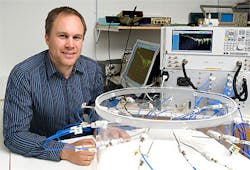
The system currently consists of 30 microwave transmitter/receivers arranged in circular fashion. The microwave radiation from the transmitters spreads out in a complex pattern across the breast and is then captured by the receivers, after which software reconstructs an image of the breast tissue in three dimensions.
Andreas Fhager, the associate professor of biomedical electromagnetics at Chalmers who developed the system, said that the 3-D images obtained from it show significantly better contrast between healthy and malignant tissue compared to x-ray-based systems. That makes it easier to detect really small tumors that may currently be obscured by healthy tissue. Unlike x-ray systems, the new system also emits negligible doses of nonionizing radiation.
With time, the Chalmers team hope to be able to use the system to not only detect the location of cancerous tissues but also to heat and destroy them with microwave radiation from the transmitters.
The underlying microwave technology is already being used in the "Strokefinder," a helmet that can distinguish between blood clots and bleeding in the brain. The Strokefinder is currently undergoing clinical trials at Sahlgrenska Hospital in Gothenburg in Sweden.
-- By Dave Wilson, Senior Editor, Vision Systems Design
