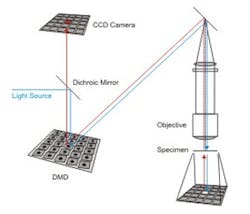
Micromirror speeds image acquisition of microscope
Confocal microscopy is routinely used for high-resolution fluorescence imaging of biological specimens. Most standard confocal systems scan a laser across a specimen and collect emitted light passing through a single pinhole to produce an optical section of the sample.
However, the sequential scanning limits the speed of image acquisition and even the fastest commercial instruments struggle to resolve the temporal dynamics of rapid cellular events.
Now, academics led by Professor Nick Hartell at Leicester University (Leicester, UK) have developed a new type of confocal microscope that promises to enable researchers to record images of cellular specimens at much higher speeds.
"Modern biological research, and modern neuroscience, depends upon the development of new technologies that allow the optical detection of biological events as they occur. Many biological events take place in the millisecond time scale and so there is a great need for new methods of detecting events at high speed and at high resolution," says Professor Hartell.
This microscope itself uses a digital micromirror which serves as both a programmable array light source and a confocal pinhole. In combination with a high-speed EMCCD camera, it can be used to obtain confocal images at speeds up to 100 frames/sec.
The researchers believe the technology will be a big help to those working in many scientific fields, including biomedical research and neuroscience.
Recently, the UK Biotechnology and Biological Sciences Research Council (BBSRC) provided funding for the research team to develop the system as a commercial product.
Hartell describes the system in detail in a recent paper -- Programmable Illumination and High-Speed, Multi-Wavelength, Confocal Microscopy Using a Digital Micromirror -- which is freely available on the PLOS One web site.
-- Dave Wilson, Senior Editor, Vision Systems Design
