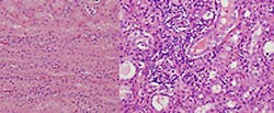
Software classifies histopathological images
For pathologists, identifying damaged or diseased tissue is a time-consuming process that involves poring over samples under a microscope.
But a collaborative research project between veterinarians and electrical engineers at Penn State University (University Park, PA, USA) has now led to the development of image processig software that could significantly speed up the process.
Working with veterinarians at the university's Animal Diagnostic Laboratory (ADL), a team led by Penn State assistant professor of electrical engineering Vishal Monga developed the automated method of classifying histopathological images. Histopathology refers to the microscopic examination of tissue in order to study the manifestations of disease.
During the course of a year, ADL's five pathologists examine more than 10,000 slides, according to Art Hattel, veterinary pathologist at ADL. To properly evaluate a slide, Hattel said a pathologist usually takes seven to 10 minutes, but the process can take as long as 20 to 25 minutes.
After initial tests showed it was feasible to use an image processing technique to classify histopathological images, Hattel received a $19,000 research grant from the Pennsylvania Department of Agriculture to initiate the project.
Working with Hattel, Monga's team developed a set of custom tools which rely on a sparse coding of the images to help with identification. The tools mimic the same methods pathologists use to classify samples by looking for distinguishing features.
Once the tools were developed, Monga said the automated solution proved to be quite accurate. It was capable of automatically categorizing the images into those that were healthy, those that were inflamed and those that exhibited necrosis with an 80- to 85-per cent success rate.
Although the new method is not being used by ADL to diagnose any real samples yet, the researchers are applying for larger grants to continue the effort in the hopes that one day it might.
Related articles from Vision Systems Design that you might also find of interest.
1. Spectroscopy aids in characterizing skin cancers
A joint US-Dutch team under the direction of Anita Mahadevan-Jansen from Vanderbilt University (Nashville, TN, USA) has developed a noninvasive probe capable of both morphological and biochemical characterization of skin cancers.
2. Software predicts cancer patient prognoses
Researchers at the Stanford University schools of engineering and medicine have developed a software-based system called Computational Pathologist, or C-Path, that can objectively and accurately assess images of breast cancer tissues to predict patient survival.
-- Dave Wilson, Senior Editor, Vision Systems Design
