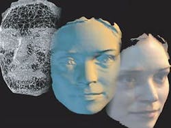
Scanning faces in 3-D helps surgical reconstruction
An event running at the Science Museum (London, UK) will allow visitors to have their faces scanned in three dimensions. The results will be used by surgeons to help improve treatment for patients with facial disfigurement.
The "Me in 3D" project is part of the Science Museum's ongoing Live Science program, where visitors can volunteer to take part in real experiments conducted by visiting scientists.
The 3-D photos will be used to create what the Science Museum says will be the largest database of 3-D facial images in the world. They will be used by surgeons from Great Ormond Street Hospital, University College Hospital, and the Eastman Dental Hospital and Institute to study patterns in face shape that could then help them improve treatment for patients with facial deformities.
"We know a lot about the bones in our faces but little is known about what makes our face the shape it is, and about the skin and muscles that make up our face. By collecting as many 3-D face photographs as we can, we will have a greater understanding of our complex faces and have greater knowledge to plan and perform the best facial surgery in the future," says Dr. Chris Abela, the Senior Craniofacial Fellow at Great Ormond Street Hospital.
"Me in 3D" will run from Jan. 1 until Apr. 10, 2012. The experiments are free and open to all visitors. No booking is required. For more information visit http://www.sciencemuseum.org.uk/mein3d.
-- By Dave Wilson, Senior Editor, Vision Systems Design
