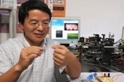
Micro-endoscope uses MEMS to detect cancer
With current camera-equipped endoscopes, once doctors spot abnormalities, they typically perform a biopsy and then send the suspicious tissue to a laboratory. But biopsy is risky and may cause bleeding and even trauma. If it is cancerous, surgeons may attempt to remove the abnormality and surrounding tissue, using either endoscopes equipped for surgery or traditional surgical methods.
Xie's endoscopes replace the cameras with infrared scanners smaller than pencil erasers. The heart of his scanner is a microelectromechanical system, or MEMS, device: A tiny motorized MEMS mirror pivots back and forth to reflect a highly focused infrared beam. By itself, the beam only strikes a period-sized dot of tissue. But the MEMS mirror allows it to move methodically back and forth, scanning a fingernail-sized piece of tissue row by row, like a lawnmower moving across a yard. The resulting image is high resolution: Xie said his scanners have achieved resolution of 10 m. Computers process the return signal from the endoscopes, transforming it into a 3-D image of the surface tissue and the tissue beneath. One scanner even produces a 360 image of all the tissue surrounding the endoscope. Doctors or other trained observers can then search the image for abnormalities or suspicious growth patterns. For more information, go to http://news.ufl.edu/2009/11/19/smart-scope/.
-- Posted by Vision Systems Design
