Researchers image hidden objects
Non-invasive optical imaging techniques, such as optical coherence tomography, are essential diagnostic tools in many disciplines, from the life sciences to nanotechnology. However, present methods are not able to image through opaque layers that scatter all the incident light. Even a very thin layer of a scattering material can appear opaque and hide any objects behind it.
Now, however, a team of researchers from the MESA+ Institute for nanotechnology at the University of Twente (Enschede, The Netherlands) in the Netherlands has succeeded in developing a method that allows non-invasive imaging of a fluorescent object that is completely hidden behind an opaque scattering layer.
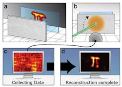
To do so, the researchers illuminated an object with laser light that had passed through a scattering layer. They then scanned the angle of incidence of the laser beam and detected the total fluorescence of the object from the front. From the detected signal, they obtained the image of the hidden object using an iterative algorithm.
As a proof of concept, they retrieved a detailed image of a fluorescent object, comparable in size (50micrometers) to a typical human cell, hidden six millimeters behind an opaque optical diffuser.
The breakthrough was published in the research journal Nature. A technical article describing the work can be found here.
Recent articles on optical coherence tomography from Vision Systems Design.
1. Imaging system gets under the skin
Researchers from Medical University Vienna (MUW; Vienna, Austria) and the Ludwig-Maximilians University (Munich, Germany) have used optical coherence tomography (OCT) to noninvasively image the network of blood vessels beneath the outer layer of skin, potentially revealing telltale signs of disease.
2. MRI system images muscles in 3-D
Researchers at the Technische Universiteit Eindhoven (TU/e; Eindhoven, The Netherlands) and the Academic Medical Center (AMC) in Amsterdam have developed a technique that allows muscle structures to be imaged in 3-D.
3. Imaging system sees bacteria behind the eardrum
A new medical imaging device invented by a research team led by University of Illinois (Champaign, IL, USA) electrical and computer engineering professor Stephen Boppart allows doctors to see biofilms behind the eardrum to better diagnose and treat chronic ear infections.
-- Dave Wilson, Senior Editor, Vision Systems Design
